Spider angioma/naevus
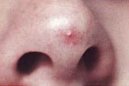
This is also called a vascular spider. It consists of a bright red, faintly pulsating lesion which has a central small blood vessel (arteriole) with slender projections radiating out from it which can be likened to spider legs.
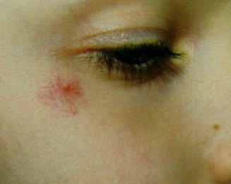
The lesions vary from a few millimetres to several centimetres in diameter.
Spider angiomas are associated with conditions in which there are high levels of the hormone oestrogen circulating, such as cirrhosis of the liver and pregnancy. However, they are also found in up to 15% of normal pre-school children and in 45% of those of school age.
The most common sites in children are the back of the hand, the forearm, the face and the ears.
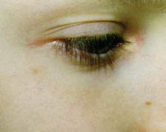
These lesions can regress on their own. They can also be treated with laser, liquid nitrogen or electrocoagulation in which the blood vessels are closed off using a low electric current.
It is hypothesized that the pulsed dye laser works in spider nevi by generating intravascular thrombi. These thrombi lodge in finer vessels, thus obliterating the system, rather than by the actual destruction of the feeding arteriole. The spider nevus is a dilated arteriole which delivers its blood into a capillary network. Histologically it has been described as containing a central ascending arteriole that ends in a thin-walled ampulla from which radiate finer arterioles. The spider nevus is exquisitely sensitive to PDL therapy because the direction of flow is from larger to finer vessels, allowing trapping of emboli. Embolization is effective only on the arterial side in end arteries and arterioles because the flow direction is from larger to smaller vessels. Embolization is not effective on the venous side of the circulation because the flow is from smaller to larger vessels, allowing small particles to wash through.